Ultrasound-guided procedures represent a significant advancement‚ evolving from optical and fluoroscopic imaging techniques to enable minimally invasive diagnostics and therapies.
These techniques‚ like UGPIV and liver biopsies‚ are becoming increasingly prevalent‚ offering enhanced precision and reduced risks for patients across various medical specialties;
What are Ultrasound-Guided Procedures?
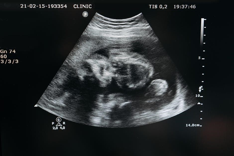
Ultrasound-guided procedures utilize real-time imaging to precisely guide instruments during various medical interventions. These techniques encompass a broad spectrum‚ including peripheral intravenous access (UGPIV)‚ liver biopsies‚ fine-needle aspirations‚ and thoracentesis.
Unlike traditional methods‚ ultrasound provides visualization of underlying structures‚ allowing healthcare professionals to navigate with increased accuracy. This minimizes the risk of complications and enhances diagnostic yield‚ ultimately improving patient outcomes. The development stems from advancements in optical and fluoroscopic imaging‚ paving the way for minimally invasive approaches.
Historical Development of Image-Guided Techniques
The evolution of image-guided techniques began with the introduction of optical imaging – like laparoscopy and cystoscopy – and fluoroscopic methods‚ such as X-ray. These innovations enabled surgeons to perform less invasive diagnostic and therapeutic procedures.
Subsequently‚ ultrasound emerged as a powerful tool‚ offering real-time visualization without ionizing radiation. This progression facilitated the development of procedures like ultrasound-guided liver biopsies and fine-needle aspirations‚ marking a significant shift towards enhanced precision and patient safety.
Applications of Ultrasound Guidance
Ultrasound guidance is broadly applied in procedures like peripheral intravenous access (UGPIV)‚ liver biopsies‚ thoracentesis‚ and fine-needle aspirations‚ improving accuracy.
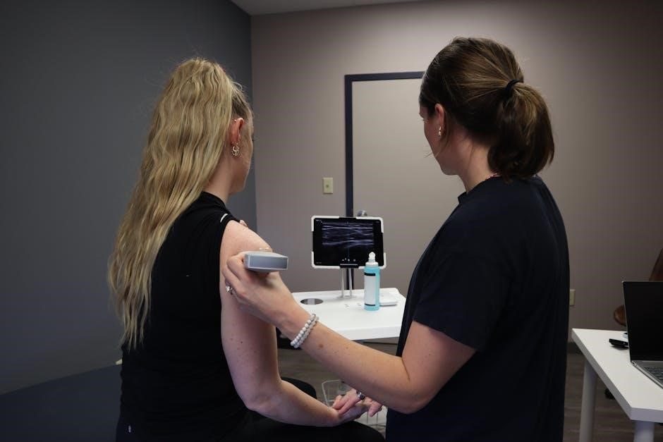
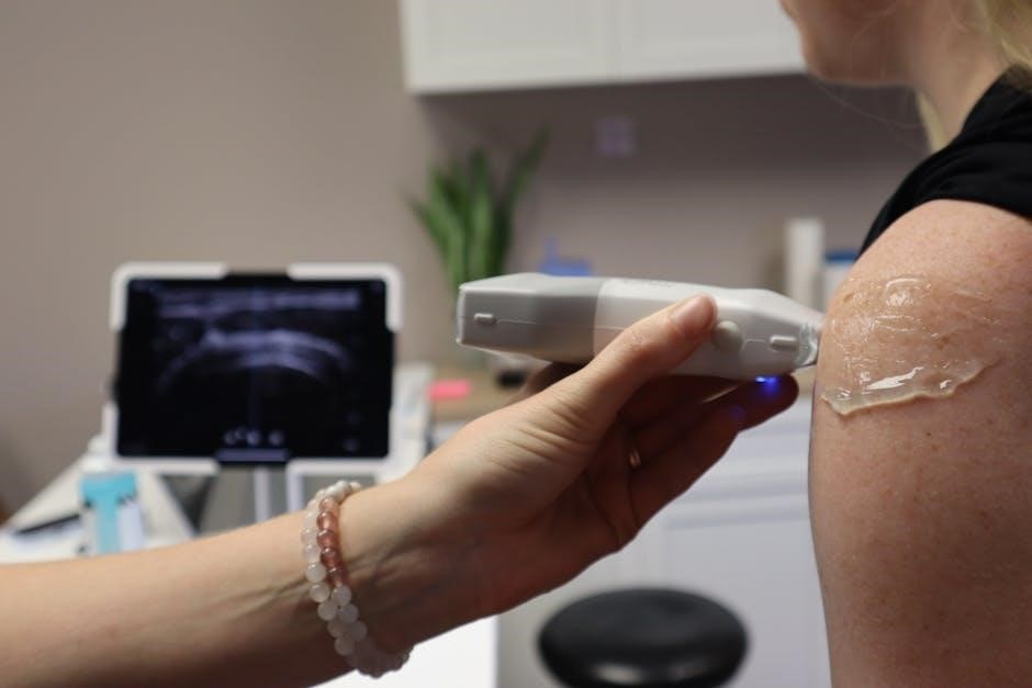
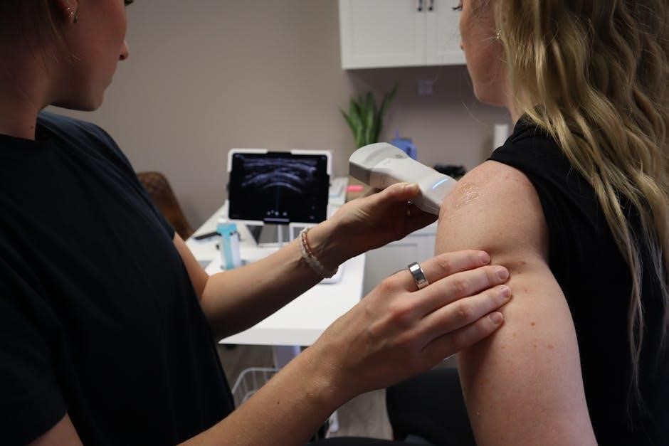
Peripheral Intravenous Access (UGPIV)
Ultrasound-guided peripheral intravenous (UGPIV) procedures are gaining prominence‚ as highlighted by recent white papers addressing associated challenges. This technique utilizes real-time imaging to locate suitable veins‚ improving success rates‚ particularly in difficult cases.
UGPIV minimizes complications like infiltration and hematoma formation‚ offering a more patient-friendly experience. Its increasing adoption reflects a shift towards enhanced precision and reduced discomfort in routine intravenous access‚ benefiting both clinicians and patients alike.
Liver Biopsy
Ultrasound-guided liver biopsies are now considered routine diagnostic procedures‚ enabling physicians to obtain small tissue samples for laboratory analysis. This technique is recommended when abnormalities are suspected‚ aiding in accurate diagnosis and treatment planning.
Specialists‚ including imaging physicians and diagnostic medical sonographers‚ utilize ultrasound to visualize the liver and guide the biopsy needle precisely. This minimizes risk and enhances the reliability of the collected sample‚ crucial for effective patient care.
Fine-Needle Aspiration
Ultrasound guidance significantly enhances the precision and safety of fine-needle aspiration (FNA) procedures. Utilizing real-time imaging‚ healthcare professionals can accurately target suspicious areas for sample collection‚ improving diagnostic yield.
Studies indicate that serious infections following ultrasound-guided procedures‚ including FNA‚ are rare. This demonstrates the procedure’s overall safety profile when performed with appropriate sterile techniques and skilled sonographer assistance‚ benefiting patient outcomes.
Thoracentesis
Ultrasound guidance is increasingly utilized during thoracentesis‚ a procedure to remove fluid from the space around the lungs. Real-time imaging allows physicians to identify optimal puncture sites‚ avoiding potential complications like pneumothorax – a collapsed lung.
Like other ultrasound-guided interventions such as biopsy and FNA‚ serious infections following thoracentesis are infrequent. This reinforces ultrasound’s role in minimizing risks and improving the safety profile of this essential diagnostic and therapeutic procedure.
Bone Structure Assessment
Ultrasound is emerging as a valuable tool for non-invasive bone assessment‚ offering a novel approach to evaluating bone structure at a microscale level. This innovative technique holds promise for assessing osteoporosis risk and monitoring treatment effectiveness without exposing patients to ionizing radiation.
Researchers are actively working to refine this technology‚ aiming to provide a readily accessible and patient-friendly method for evaluating bone health and guiding clinical decisions.
Technological Advancements
Robotics‚ like those developed by Mendaera‚ are receiving FDA clearance‚ enhancing ultrasound-guided procedures‚ while new non-invasive bone assessment techniques are emerging.
Robotics in Ultrasound-Guided Procedures
Robotics are increasingly integrated into ultrasound-guided procedures‚ offering enhanced precision and control for healthcare professionals. Companies like Mendaera are pioneering this field‚ receiving FDA 510(k) clearance for robotic systems designed for universal deployment in medicine.
This technological leap promises to improve accuracy during interventions like biopsies and fine-needle aspirations‚ potentially minimizing operator variability and expanding access to advanced imaging techniques. The goal is to create more consistent and reliable outcomes for patients undergoing these procedures.
FDA Clearance and New Technologies
Recent advancements in ultrasound guidance are rapidly gaining FDA clearance‚ signifying their safety and efficacy. Mendaera’s robotic system exemplifies this progress‚ receiving 510(k) clearance for broader medical application.
Furthermore‚ non-invasive bone assessment techniques utilizing ultrasound are emerging‚ offering potential for early osteoporosis detection and treatment monitoring. These innovations highlight a shift towards less invasive diagnostic methods and improved patient care through cutting-edge technology.
Non-Invasive Bone Assessment Techniques
Emerging ultrasound techniques provide a revolutionary‚ non-invasive method for evaluating bone structure at a microscopic level. Researchers are actively refining these methods to accurately assess osteoporosis risk and monitor the effectiveness of various treatments.
This innovative approach eliminates the need for more invasive procedures‚ offering a comfortable and accessible option for patients and potentially improving early detection rates.
Benefits of Ultrasound Guidance
Ultrasound guidance delivers a minimally invasive approach‚ boosting accuracy and precision while significantly reducing complication risks for patients undergoing procedures.
Minimally Invasive Approach
Ultrasound guidance facilitates procedures with smaller incisions compared to traditional surgical methods‚ leading to less tissue trauma and reduced postoperative pain. This approach minimizes scarring and promotes faster recovery times for patients.
Techniques like UGPIV and liver biopsies‚ when ultrasound-guided‚ require only a small needle insertion‚ avoiding larger surgical openings. This translates to decreased risk of infection‚ reduced blood loss‚ and a quicker return to normal activities‚ enhancing patient comfort and overall outcomes.
Increased Accuracy and Precision
Ultrasound guidance provides real-time visualization of internal structures‚ allowing healthcare professionals to precisely target specific areas during procedures like fine-needle aspiration or thoracentesis. This visual feedback significantly improves the accuracy of needle placement‚ reducing the chance of misdiagnosis or damage to surrounding tissues.
By directly visualizing the target‚ clinicians can navigate around vessels and organs‚ ensuring optimal sample acquisition or fluid drainage‚ ultimately leading to more reliable results and improved patient care.
Reduced Risk of Complications
Ultrasound guidance demonstrably lowers the incidence of complications associated with invasive procedures. Studies indicate that serious infections following biopsies‚ fine-needle aspirations‚ and thoracentesis are rare when ultrasound is utilized.
The real-time imaging minimizes the risk of hitting blood vessels or other critical structures‚ thereby decreasing the likelihood of bleeding and hematoma formation. This translates to safer procedures and faster recovery times for patients.
Potential Risks and Complications
Ultrasound-guided procedures‚ while generally safe‚ carry potential risks including infection‚ though rare‚ and the possibility of bleeding or hematoma development post-procedure.
Infection Rates
Infection following ultrasound-guided procedures‚ such as biopsies‚ fine-needle aspirations‚ and thoracentesis‚ is considered a relatively uncommon complication. Studies indicate that serious infections are rarely observed in patients undergoing these image-guided interventions.
However‚ as with any invasive procedure‚ maintaining strict sterile techniques during the process is crucial to minimize the risk of introducing pathogens. Vigilant monitoring for signs of infection post-procedure remains essential for prompt diagnosis and treatment‚ ensuring optimal patient outcomes.
Bleeding and Hematoma Formation
Bleeding and subsequent hematoma formation represent potential‚ though generally minor‚ complications associated with ultrasound-guided procedures. The risk is influenced by factors like the specific procedure performed‚ patient’s coagulation status‚ and operator skill.
Real-time ultrasound imaging allows for visualization of blood vessels‚ aiding in precise needle placement to minimize vascular injury. Post-procedure compression can further reduce the likelihood of hematoma development‚ contributing to improved patient comfort and recovery.
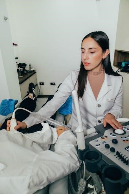
The Role of Healthcare Professionals
Imaging physicians‚ diagnostic medical sonographers‚ and surgeons collaborate in ultrasound-guided procedures‚ utilizing their expertise for accurate diagnoses and effective treatments.
Imaging Physicians
Imaging physicians play a crucial role in ultrasound-guided procedures‚ possessing specialized training in interpreting ultrasound images and guiding interventions. They are responsible for accurately visualizing anatomical structures‚ identifying potential abnormalities‚ and ensuring precise needle placement during biopsies or aspirations.
Their expertise extends to assessing the risks and benefits of each procedure‚ collaborating with surgeons and sonographers‚ and overseeing the overall imaging process to optimize patient outcomes. They are key to accurate diagnoses.
Diagnostic Medical Sonographers
Diagnostic Medical Sonographers are integral to ultrasound-guided procedures‚ operating the ultrasound equipment and acquiring real-time images under the supervision of a physician. They possess a deep understanding of ultrasound physics and anatomy‚ skillfully maneuvering the transducer to visualize target areas.
Sonographers assist in guiding needles‚ documenting findings‚ and ensuring image quality throughout the procedure‚ contributing significantly to accuracy and patient safety. Their role is vital for successful interventions.
Surgeons Utilizing Ultrasound
Surgeons are increasingly integrating ultrasound guidance into their practice‚ leveraging real-time imaging for enhanced precision during both diagnostic and therapeutic interventions. This approach minimizes invasiveness‚ reducing patient trauma and accelerating recovery times.
From liver biopsies to fine-needle aspirations‚ surgeons rely on ultrasound to visualize structures‚ guide instruments‚ and confirm accurate placement‚ ultimately improving procedural outcomes and patient care.
Ultrasound Equipment and Techniques
Ultrasound employs various transducers and Doppler applications for real-time imaging‚ crucial for guiding procedures and visualizing anatomical structures with precision.
Types of Ultrasound Transducers
Ultrasound transducers convert electrical energy into sound waves and vice versa‚ differing significantly based on frequency and application. Linear transducers are ideal for superficial structures‚ providing high-resolution images for UGPIV and vascular access.
Curved transducers offer a wider field of view‚ suitable for deep organ imaging like liver biopsies. Phased array transducers‚ often used for cardiac imaging‚ can be maneuvered within vessels. Selecting the appropriate transducer is paramount for optimal image quality and procedural success‚ influencing diagnostic accuracy.
Real-Time Imaging
Real-time imaging is the cornerstone of ultrasound-guided procedures‚ allowing healthcare professionals to visualize anatomy and guide instruments dynamically. This capability is crucial for procedures like thoracentesis and fine-needle aspiration‚ ensuring accurate needle placement and minimizing risk.
The continuous visual feedback provided by real-time ultrasound enhances precision‚ avoids critical structures‚ and confirms successful tissue sampling‚ ultimately improving patient safety and procedural outcomes across diverse clinical applications.
Doppler Ultrasound Applications
Doppler ultrasound expands the utility of ultrasound-guided procedures by visualizing blood flow‚ offering critical information beyond anatomical structures. This is particularly valuable during peripheral intravenous access (UGPIV)‚ helping identify suitable veins and assess vessel patency.
Furthermore‚ Doppler aids in evaluating vascularity within biopsied tissues and detecting potential bleeding or hematoma formation post-procedure‚ contributing to enhanced monitoring and improved patient care.
Future Trends in Ultrasound Guidance
Artificial intelligence (AI) integration and enhanced imaging resolution are poised to revolutionize ultrasound guidance‚ promising greater accuracy and efficiency in procedures.
Artificial Intelligence Integration
Artificial intelligence (AI) is rapidly transforming ultrasound-guided procedures‚ offering potential for automated image analysis and enhanced decision-making capabilities. AI algorithms can assist in real-time needle guidance‚ improving precision and reducing operator dependence.
Furthermore‚ AI can aid in identifying subtle anatomical variations and potential complications‚ leading to safer and more effective interventions. Machine learning models are being developed to predict optimal biopsy sites and personalize treatment strategies based on individual patient characteristics‚ ultimately improving outcomes.
Enhanced Imaging Resolution
Future trends in ultrasound guidance heavily focus on achieving significantly enhanced imaging resolution. This involves developing new transducer technologies and signal processing techniques to visualize anatomical structures with greater clarity and detail.
Higher resolution imaging allows for more accurate needle placement‚ improved visualization of small lesions‚ and better differentiation between tissues. These advancements are crucial for complex procedures‚ ultimately leading to increased diagnostic accuracy and therapeutic efficacy for patients undergoing ultrasound-guided interventions.
Training and Certification
Sonography training programs and continuing medical education are vital for healthcare professionals mastering ultrasound guidance‚ ensuring competency and safe practice.
Sonography Training Programs
Comprehensive sonography training programs are foundational for developing skilled professionals in ultrasound-guided procedures. These programs typically encompass detailed anatomical study‚ physics of ultrasound‚ and hands-on clinical experience.
Curricula focus on image acquisition‚ interpretation‚ and procedural techniques specific to various applications like UGPIV‚ biopsies‚ and thoracentesis. Accreditation by organizations ensures program quality and adherence to established standards. Successful completion prepares sonographers to collaborate effectively with imaging physicians and surgeons.
Continuing Medical Education
Continuing Medical Education (CME) is crucial for healthcare professionals utilizing ultrasound-guided procedures‚ given the rapid technological advancements in the field. CME courses cover new techniques‚ like non-invasive bone assessment‚ and updates on FDA-cleared technologies‚ such as robotic assistance.
These programs ensure practitioners maintain competency in image interpretation‚ procedural skills‚ and safety protocols‚ ultimately enhancing patient care and minimizing potential complications.
Cost-Effectiveness of Ultrasound Guidance
Ultrasound guidance demonstrably lowers healthcare expenses through reduced hospital stays and overall treatment costs‚ making it a financially sound medical approach.
Reduced Hospital Stay
Ultrasound-guided procedures frequently allow for shorter hospitalizations compared to traditional‚ open surgical methods. The minimally invasive nature of techniques like UGPIV‚ liver biopsies‚ and thoracentesis contributes to faster patient recovery times.
This reduction in length of stay directly translates to lower healthcare costs‚ freeing up valuable hospital resources and improving patient throughput. Precise targeting and minimized trauma‚ facilitated by ultrasound‚ lessen post-operative complications‚ further supporting quicker discharge planning and improved patient outcomes.
Lower Overall Treatment Costs
Ultrasound guidance contributes to significant cost savings within healthcare systems. By enabling minimally invasive procedures like fine-needle aspiration and liver biopsies‚ the need for extensive surgical interventions is often avoided.
This translates to reduced anesthesia fees‚ shorter hospital stays – as previously noted – and fewer post-operative complications requiring additional treatment. The increased accuracy also minimizes repeat procedures‚ further lowering the overall treatment costs for patients and healthcare providers alike.

Ethical Considerations
Ultrasound-guided procedures demand strict adherence to ethical principles‚ prioritizing informed patient consent and ensuring accurate image interpretation for optimal‚ responsible care.
Patient Consent and Information
Obtaining informed consent is paramount before any ultrasound-guided procedure. Patients must receive comprehensive information regarding the procedure’s purpose‚ potential benefits‚ and inherent risks‚ including infection or bleeding.
Clear communication about alternative diagnostic options is also crucial‚ empowering patients to make autonomous decisions. Ensuring patients understand the image interpretation process and potential outcomes fosters trust and shared responsibility in their healthcare journey. Thorough documentation of the consent process is essential for ethical and legal compliance.
Image Interpretation and Accuracy
Accurate image interpretation is fundamental to the success of ultrasound-guided procedures. Skilled imaging physicians and diagnostic medical sonographers must possess expertise in recognizing normal anatomy and pathological findings.
Variations in technique and equipment can influence image quality‚ necessitating continuous training and quality control measures. Maintaining objectivity and minimizing interpretive errors are vital for patient safety and optimal treatment planning‚ ensuring reliable diagnostic outcomes.
Research and Development
Ongoing clinical trials are actively exploring new applications of ultrasound guidance‚ alongside advancements in artificial intelligence integration and enhanced imaging resolution.
Ongoing Clinical Trials
Numerous clinical trials are currently investigating the expanded role of ultrasound-guided procedures across diverse medical fields. Research focuses on optimizing techniques for liver biopsies‚ fine-needle aspirations‚ and thoracentesis‚ aiming to minimize complications like infection and bleeding.
Furthermore‚ studies are evaluating the efficacy of robotic assistance in enhancing precision and accessibility. Investigations also explore the potential of non-invasive bone assessment using advanced ultrasound techniques‚ particularly for osteoporosis risk evaluation and treatment monitoring.
New Applications Being Explored
Researchers are actively investigating novel applications for ultrasound guidance beyond established procedures. This includes exploring its utility in targeted drug delivery‚ enhancing the accuracy of nerve blocks for pain management‚ and improving the visualization of subtle anatomical structures during complex surgeries.
The development of artificial intelligence (AI) integration promises to further expand possibilities‚ potentially automating image interpretation and optimizing procedural workflows. Non-invasive bone assessment continues to be refined‚ offering a promising alternative to traditional methods.